In this article, we will explore the importance of Magnetic Resonance Imaging (MRI) in understanding post-surgical changes. After undergoing surgery, patients often experience various changes in their bodies that can be challenging to assess through physical examination alone. MRI plays a crucial role in providing detailed insights into these changes, allowing healthcare professionals to make informed decisions regarding patient care and treatment.
Brief explanation of post-surgical changes
Post-surgical changes refer to the alterations that occur in the body following a surgical procedure. These changes can manifest in different ways, such as scarring, inflammation, edema, hematoma, seroma, or implant-related modifications. Understanding these changes is essential for monitoring the healing process, detecting complications, and assessing the effectiveness of treatment.
Importance of MRI in understanding these changes
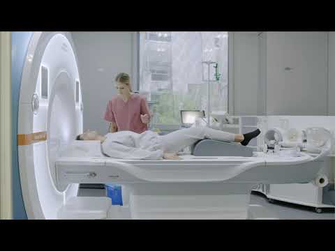
MRI is a powerful imaging technique that utilizes a combination of magnetic fields and radio waves to create detailed images of the body’s internal structures. Unlike other imaging techniques, such as X-rays or CT scans, MRI provides a non-invasive and radiation-free method to visualize soft tissues, making it particularly valuable in assessing post-surgical changes.
MRI allows healthcare professionals to visualize and evaluate the extent of these changes, providing valuable information about the underlying causes and effects. By accurately interpreting MRI findings, healthcare professionals can make informed decisions regarding patient management and treatment strategies.
In the following sections, we will delve deeper into the understanding of MRI technology, its advantages over other imaging techniques, and the common post-surgical changes that can be detected through MRI. We will also explore the role of MRI in post-surgical follow-up and present case studies to illustrate its practical applications. Finally, we will conclude by emphasizing the importance of utilizing MRI for a better understanding and management of post-surgical outcomes.
Understanding MRI
MRI, which stands for Magnetic Resonance Imaging, is a powerful diagnostic tool that uses a combination of magnetic fields and radio waves to create detailed images of the body’s internal structures. It is widely used in the medical field to aid in the diagnosis and monitoring of various conditions, including post-surgical changes. Understanding how MRI works and its advantages over other imaging techniques is crucial in appreciating its role in decoding these changes.
Explanation of MRI technology
MRI technology relies on the principle of nuclear magnetic resonance, which involves the interaction of atomic nuclei with magnetic fields. The human body is composed of atoms, and certain atoms, such as hydrogen, have a property called spin. When placed in a strong magnetic field, these atoms align themselves with the field.
How MRI works to capture detailed images
To capture images, MRI machines generate a strong magnetic field that aligns the hydrogen atoms in the body. Radio waves are then applied, causing the atoms to emit signals. These signals are detected by the MRI machine and processed to create detailed cross-sectional images of the body. By manipulating the magnetic field and radio waves, different types of tissue can be distinguished, providing valuable information about the structures being imaged.
Advantages of MRI over other imaging techniques
MRI offers several advantages over other imaging techniques, making it particularly useful in decoding post-surgical changes. Firstly, MRI does not use ionizing radiation, unlike X-rays and CT scans, making it safer for patients, especially those who require repeated imaging. Secondly, MRI provides excellent soft tissue contrast, allowing for the visualization of subtle changes in the body. This is particularly important when assessing post-surgical changes, as it enables the detection of abnormalities that may not be visible on other imaging modalities. Lastly, MRI is a non-invasive procedure that does not require the use of contrast agents in most cases, minimizing the risk of adverse reactions.
In summary, MRI is a valuable tool in understanding post-surgical changes. Its ability to capture detailed images, excellent soft tissue contrast, and non-invasive nature make it an ideal choice for decoding these changes. By utilizing MRI, healthcare professionals can gain valuable insights into the effects of surgery, monitor healing progress, and assess the effectiveness of treatments. Patients and healthcare professionals alike should embrace the use of MRI to enhance their understanding and management of post-surgical outcomes.
Common Post Surgical Changes Detected by MRI
Post-surgical changes are a natural part of the healing process after undergoing a surgical procedure. These changes can be complex and varied, making it crucial to have a comprehensive understanding of them. Magnetic Resonance Imaging (MRI) plays a vital role in detecting and interpreting these changes, providing valuable insights for both patients and healthcare professionals.
Scarring and Fibrosis
Causes and Effects:
- Scarring and fibrosis occur as a result of the body’s natural response to tissue injury during surgery.
- The formation of scar tissue is a normal part of the healing process, but excessive scarring can lead to complications.
- Fibrosis refers to the excessive accumulation of connective tissue, which can cause stiffness and restricted movement.
MRI Findings and Interpretation:
- MRI can accurately visualize and assess the extent of scarring and fibrosis.
- It provides detailed images that help identify the location, size, and characteristics of scar tissue.
- By evaluating the MRI findings, healthcare professionals can determine the impact of scarring and fibrosis on the patient’s recovery and plan appropriate interventions.
Edema and Inflammation
Causes and Effects:
- Edema, or swelling, occurs due to the accumulation of fluid in the tissues surrounding the surgical site.
- Inflammation is the body’s response to tissue injury, characterized by redness, heat, swelling, and pain.
- Excessive edema and inflammation can impede the healing process and prolong recovery.
MRI Findings and Interpretation:
- MRI can detect and quantify the extent of edema and inflammation.
- It provides valuable information about the distribution and severity of these changes.
- By assessing the MRI findings, healthcare professionals can determine the effectiveness of anti-inflammatory treatments and adjust the patient’s management plan accordingly.
Hematomas and Seromas
Causes and Effects:
- Hematomas are collections of blood that accumulate in the surgical area, often caused by blood vessel damage during surgery.
- Seromas are fluid collections that form due to the accumulation of lymphatic fluid.
MRI Findings and Interpretation:
- MRI is highly sensitive in detecting and characterizing hematomas and seromas.
- It can differentiate between active bleeding and older blood collections.
- By evaluating the MRI findings, healthcare professionals can determine the need for intervention, such as drainage or surgical removal.
Implant-Related Changes
Causes and Effects:
- Implant-related changes refer to alterations in the position, integrity, or function of surgical implants.
- These changes can occur due to implant migration, rupture, or other complications.
MRI Findings and Interpretation:
- MRI is an excellent tool for evaluating implant-related changes.
- It can visualize the implants and surrounding tissues, providing detailed information about their condition.
- By assessing the MRI findings, healthcare professionals can identify implant-related issues early on and plan appropriate interventions.
Understanding the common post-surgical changes detected by MRI is crucial for effective patient management. MRI offers a non-invasive and detailed assessment of these changes, allowing healthcare professionals to make informed decisions regarding treatment and follow-up care. By utilizing MRI, patients and healthcare professionals can work together to ensure optimal outcomes and a smoother recovery process.
Role of MRI in Post Surgical Follow-up
After undergoing surgery, it is crucial to closely monitor the healing process and detect any potential complications. Magnetic Resonance Imaging (MRI) plays a vital role in post-surgical follow-up, providing detailed information about the changes occurring within the body. Let’s explore the various ways in which MRI contributes to the management of post-surgical outcomes.
Early detection of complications
One of the primary benefits of MRI in post-surgical follow-up is its ability to detect complications at an early stage. Complications such as infections, fluid collections, or abnormal tissue growth can be identified through MRI scans. By detecting these issues early on, healthcare professionals can intervene promptly and prevent further complications.
Monitoring healing progress
MRI allows healthcare professionals to monitor the healing progress of surgical sites. By capturing detailed images, MRI can reveal the formation of scar tissue and fibrosis, which are common post-surgical changes. These changes can impact the overall outcome of the surgery and may require additional interventions. Regular MRI scans enable healthcare professionals to assess the healing process and make informed decisions regarding further treatment.
Assessing treatment effectiveness
In some cases, post-surgical treatments such as chemotherapy or radiation therapy may be necessary. MRI plays a crucial role in assessing the effectiveness of these treatments. By comparing pre and post-treatment MRI scans, healthcare professionals can evaluate the response of tumors or other abnormalities to the treatment. This information helps in determining whether the treatment is working as expected or if any adjustments are required.
MRI also aids in evaluating the success of implant-related surgeries. Whether it is a joint replacement or breast augmentation, MRI can provide detailed images of the implants and surrounding tissues. This allows healthcare professionals to assess the position, integrity, and potential complications associated with the implants.
Limitations of MRI in post-surgical follow-up
While MRI is a powerful tool in post-surgical follow-up, it does have some limitations. Metallic implants or objects within the body can cause artifacts in the MRI images, making interpretation challenging. Additionally, MRI scans can be time-consuming and expensive compared to other imaging techniques. However, the benefits of MRI in post-surgical follow-up often outweigh these limitations, making it a valuable tool in patient care.
MRI plays a crucial role in post-surgical follow-up, providing valuable insights into the changes occurring within the body. It enables early detection of complications, monitoring of healing progress, and assessment of treatment effectiveness. By utilizing MRI, healthcare professionals can make informed decisions, ensure optimal patient care, and improve post-surgical outcomes. It is essential for both patients and healthcare professionals to recognize the importance of MRI in post-surgical follow-up and utilize this powerful imaging technique for better understanding and management of post-surgical outcomes.
Case Studies
In this section, we will explore two case studies that highlight the significance of MRI in understanding post-surgical changes. These examples demonstrate how MRI imaging can provide valuable insights into the healing process and help healthcare professionals make informed decisions regarding treatment and follow-up care.
Example 1: MRI findings after knee surgery
Description of the case
In this case study, a 45-year-old patient underwent knee surgery to repair a torn meniscus. Following the procedure, the patient experienced persistent pain and limited range of motion. To assess the post-surgical changes, an MRI scan was performed.
MRI images and interpretation
The MRI images revealed the presence of scarring and fibrosis in the surgical site. Scarring occurs as a natural part of the healing process, but excessive scarring can lead to complications such as restricted movement and chronic pain. The MRI findings allowed the healthcare team to visualize the extent of scarring and determine the appropriate course of action. In this case, physical therapy and targeted interventions were recommended to address the scarring and improve the patient’s mobility.
Example 2: MRI findings after breast augmentation
Description of the case
In this case study, a 30-year-old woman underwent breast augmentation surgery to enhance her breast size and shape. Several months after the procedure, the patient noticed asymmetry and discomfort in her breasts. An MRI scan was performed to evaluate the post-surgical changes and identify the underlying cause.
MRI images and interpretation
The MRI images revealed the presence of a hematoma in one of the breasts. A hematoma is a collection of blood that can occur as a result of surgery. It can cause pain, swelling, and distortion of the surgical site. The MRI findings confirmed the presence of a hematoma, allowing the healthcare team to promptly address the issue. In this case, a drainage procedure was performed to remove the hematoma and alleviate the patient’s symptoms.
These case studies demonstrate the crucial role of MRI in post-surgical follow-up. By providing detailed and accurate images, MRI enables healthcare professionals to detect complications early, monitor the healing progress, and assess the effectiveness of treatments. This information is invaluable in guiding patient care and ensuring optimal outcomes.
In conclusion, MRI plays a vital role in understanding post-surgical changes. Through its advanced imaging technology, MRI provides healthcare professionals with detailed insights into the healing process and helps them make informed decisions regarding treatment and follow-up care. The case studies presented in this article highlight the importance of MRI in detecting and interpreting post-surgical changes, enabling timely interventions and improved patient outcomes. It is essential for both patients and healthcare professionals to recognize the value of MRI in decoding post-surgical outcomes and utilize this powerful tool for better understanding and management of post-surgical changes.
